Macular Degeneration
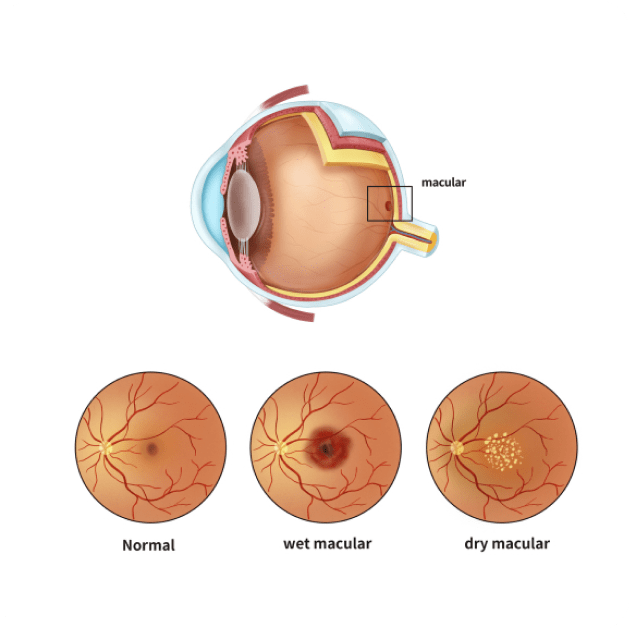
The macula is located in the center of the retina at the back of your eye. Your macula is responsible for providing sharp central vision, allowing you to see objects with clarity and detail. Age-Related Macular Degeneration (AMD) occurs when the macula gradually deteriorates over time, leading to a gradual loss of detailed central vision.
There are two main types of AMD:
Dry AMD
In Dry AMD, yellowish deposits called drusen accumulate beneath the macula causing gradual thinning of the retinal tissue over time. The progression of dry AMD is, however, very slow and may take many years to reach to an advanced stage.
Wet AMD
In wet AMD, abnormal blood vessels begin to grow beneath the retina. These new blood vessels are fragile and can leak blood and fluid, causing damage to the macula and progressive vision loss. Wet AMD tends to progress more rapidly than Dry AMD and has the potential to cause more severe central vision loss without treatment.
Symptoms of AMD will vary on the type and stage of degeneration. Early AMD may not cause any symptoms initially.
As AMD progresses you may slowly notice:
- Difficulty with tasks that require clear central vision such as reading, driving, watching TV, and recognising faces
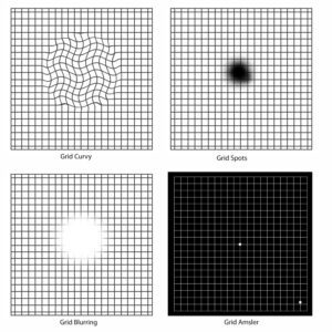
- Straight lines may appear wavy or distorted
- Dark spots or patchy central vision
It’s important to note that these symptoms typically affect the central part of the visual field, while peripheral vision remains intact
The best way for a patient to monitor macular degeneration at home is by using an Amsler grid. If any significant change is noticed on the Amsler grid it can suggest progression of macular degeneration and you should see your retina specialist urgently.

There is currently no cure for dry AMD. Early diagnosis, regular eye examinations, and reducing AMD risk factors can help slow retinal disease progression. These include:
- A diet rich in antioxidants like green leafy vegetables (spinach, broccoli), fruits rich in vitamin C (citrus fruits, berries, melons), fish and nuts
- Avoid smoking
- There is evidence from large studies (AERDS) that nutritional supplements specifically for macula health can slow the progression of retinal disease.
All the dietary advice needs to be taken after consulting with your GP or nutritionist
Retinal disease treatment for wet AMD aims to stop new blood vessels from growing and damaging the macula. The Main retinal disease treatment options for wet AMD is
Anti-VEGF Injections:
Anti-VEGF is a medication that inhibits the growth of abnormal blood vessels and reduces leakage and swelling under the macula. The medication is injected into the affected eye and is usually performed in the office under local anesthetic drops.
For more information on Intravitreal injections please click here
There is no exact cause of AMD, however, there are certain factors that can contribute to its development. These include age, genetics, smoking, nutrition, and cardiovascular disease.

AMD is usually diagnosed during a comprehensive eye examination. Initial testing includes vision assessment and a thorough retinal examination. If there are concerns about your macula, you may require some additional specialised tests that focus on the structure and function of your macula. These tests include:
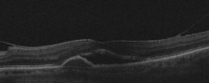
Optical Coherence Tomography (OCT) and OCT Angiography
A non-invasive imaging scan that provides detailed cross-sectional images of the macula. It is used to detect and monitor any macular abnormalities.
Retinal Photography and Autofluorescence
Using a specialized wide-view camera that takes images of your retina to detect and monitor macular and retinal conditions.
Fluorescein Angiography / Indocyanine Green Angiography
A diagnostic procedure used to examine the blood flow in the retina. It involves injecting a special dye called Fluorescein or ICG into a vein in your arm, then taking a series of photographs as the dye circulates through the blood vessels in the retina. The test identifies areas of leaking or abnormal blood vessels, blockages, swelling or other macula changes.
