What is a Vitrectomy?
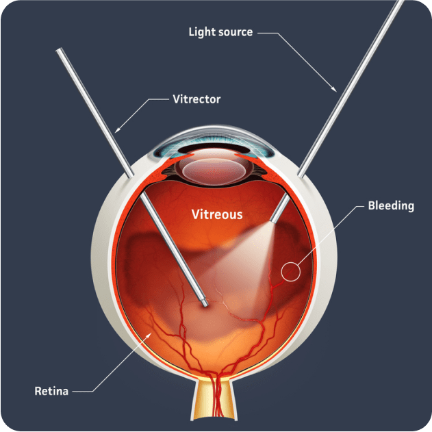
Vitreous humour is a transparent gel-like substance that fills the eye cavity. A vitrectomy is a surgical procedure that involves removing the vitreous humour from the eye and replacing it with a different solution.
The most common conditions that require a vitrectomy include:
Epiretinal Membrane (ERM)
An epiretinal membrane (ERM), also known as macular pucker or cellophane maculopathy, is a thin layer of scar tissue that develops on the surface of the retina at the back of the eye.

ERM can develop due to various factors, including age-related changes, previous eye surgeries, inflammation, or eye injuries.
Mild cases of ERM may not cause noticeable symptoms. More advanced membranes can pull on the macula, the central part of the retina, and cause distorted or wavy vision, difficulty reading or decreased central vision.
Treatment for ERM may not be necessary if the symptoms are mild and do not significantly affect vision. Ongoing monitoring is recommended as ERM can worsen over time, especially with age.
In cases where vision is affected, the sole available treatment option is surgery. This involves performing a vitrectomy and removing the epiretinal membrane.
Macular hole
A Macular Hole is a small, round hole that forms right at the centre of the macula.
The most common cause of a macular hole is a natural aging process where the vitreous gel pulls away from the retina. Occasionally the vitreous can pull on the central area of the macula and result in the formation of a macular hole.
Macular Holes can go through different stages as they develop, from partial to full thickness holes, and their severity can increase over time.

Treatment of a macular hole often involves vitrectomy surgery to remove the vitreous gel and release the traction. Long-acting gas is often injected following this to keep the macula and retina attached post-operatively.
Retinal detachment

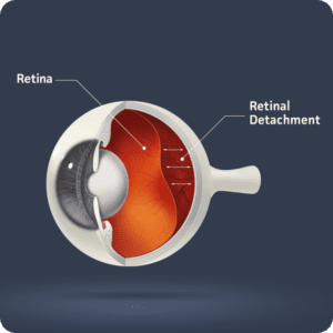
A retinal detachment is a serious eye condition where the retina becomes separated from its underlying support layers at the back of the eye.
There are several factors that can cause a retinal detachment such as traction between the vitreous gel and retina, trauma, previous eye surgeries, inflammation, retinal tears or a high degree of myopia.
This can cause sudden and significant changes in vision including:
- Floaters and Flashes Click here
- A curtain-like shadow obstructing part of the visual field
- Sudden blurry or significant loss of vision
A retinal detachment is considered a medical emergency and immediate surgical treatment is usually required. Usually, a vitrectomy is required to remove the vitreous gel and a gas bubble or silicone oil may be injected to keep the retina in place while it heals. Less common surgical options include cryotherapy or a scleral buckle.
A vitrectomy is a surgical procedure performed in a hospital. The procedure is usually performed under a local or general anaesthetic, so fasting may be required.
The eye surgeon makes very small incisions in the sclera (the white of the eye), usually less than 1mm in size, to remove the vitreous and repair the eye condition being treated. The vitreous is then replaced by injecting gas, silicone oil, or saline to stabilise the retina. The incisions are so small that they often ‘self-seal’ and do not require stitches. Antibiotic ointment and a patch is then placed over the eye.
Following the procedure, your eye may be red and sore, and your vision may be blurry. If a gas bubble has been injected, blurry vision can persist for 2-4 weeks until the gas bubble dissipates and the natural fluids in your eye replenish. During this time you must avoid air travel and exposure to high altitudes.
Specific postoperative instructions and recommendations will vary on the type of surgery performed. Your surgeon will provide you with detailed instructions and postoperative care
