What is Retinal Vein Occlusion?
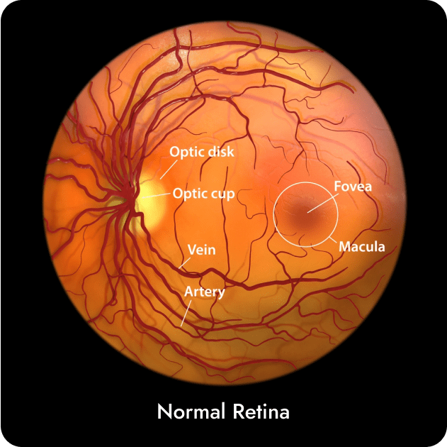
Retinal Vein Occlusion (RVO) is a condition where the veins carrying blood away from the retina, located at the back of the eye, become blocked. This blockage causes blood and fluid to leak into the retina, leading to visual problems.
The main types of RVO:
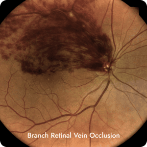
Branch Retinal Vein Occlusion (BRVO)
BRVO occurs when one or more branches of the central retinal vein are blocked. This affects a smaller area of the retina compared to CRVO. However, it can cause swelling in the macula and affect central vision. Vision loss in BRVO is usually not as severe as in CRVO.

Central Retinal Vein Occlusion (CRVO)
In CRVO, the main central retinal vein, which drains blood from the entire retina, becomes blocked. This leads to poor blood flow throughout the retina and can cause severe vision loss.
The type of retinal vein occlusion determines the extent of visual impairment and the appropriate retinal disease treatment options. It is important to consult with a retina eye specialist for a proper diagnosis and management of the condition.
RVO can affect both men and women, typically occurring in individuals above the age of 60. However, there are several underlying conditions and lifestyle factors that can contribute to its development including:
- Cardiovascular disease, including high blood pressure and cholesterol
- Being overweight, poor diet, and exercise
- Smoking
- Stress
- Diabetes
- Blood clotting disorders
RVO is diagnosed through a combination of clinical evaluations and specialised tests including:
Dilated eye examination
This involves using eye drops to enlarge your pupils and allows the eye specialist to thoroughly examine the retina at the back of your eye.
Optical Coherence Tomography (OCT) and OCT Angiography
A non-invasive imaging scan that provides detailed cross-sectional images of the retina and aids in the diagnosis and treatment of vascular eye disease.
Fluorescein Angiography
A diagnostic procedure used to examine the blood flow in the retina. It involves injecting a special dye called Fluorescein into a vein in your arm, then taking a series of photographs as the dye circulates through the blood vessels in the retina. This helps to evaluate the blood flow and detect any blockages or abnormalities in the retinal circulation.
Systemic Examination
Since RVO can be associated with underlying systemic conditions, a complete systemic evaluation is important. This may involve assessing blood pressure, examining the carotid arteries (using imaging techniques such as ultrasound), and evaluating the patient’s medical history for risk factors like hypertension, diabetes, or cardiac disorders. All the cardiovascular risk factors must be optimised with the help of your GP.
- Sudden blurry, patchy or total loss of vision in one eye only
- New or sudden increase in floaters
- Eye pain or headache (in rare cases)
The treatment of RVO depends on the severity of the condition and its impact on vision.
In mild cases, where vision is not significantly affected, close monitoring and lifestyle changes for improved cardiovascular health may be recommended.
For more severe cases, treatment options include:
Pharmacological treatment
These include medications to reduce inflammation, improve blood circulation, prevent blood clot formation, or reduce eye pressure.
Intravitreal injections
An injection of medication into the eye to treat leaking blood vessels and reduce swelling.
To read more about Intravitreal injections please click here.
Retinal laser treatment
Laser treatment is used to seal off any leaking blood vessels, reduce swelling and inhibit abnormal blood vessel growth.
To read more about Retinal laser treatment please click here
