If you have been referred to a Retina Specialist for a macular assessment, you may be wondering what happens at your appointment?
On your first visit to an Ophthalmologist, you will require a comprehensive examination of your eyes to assess their overall health and detect any potential problems. Your eye has several components and a thorough assessment involves more than just a vision check.
A full eye assessment usually consists of several tests performed by your Retina Specialist and their clinical team. You may have had some of these tests performed at your optometrist or previous eye doctor; these tests usually need to be repeated using specialised equipment, so we have current and accurate results on the day of your appointment.
Macular Health Importance
Your macula is located in the centre of the retina at the back of your eye. Your macula is responsible for providing detailed central vision. Maintaining good macular health is essential for overall visual function, allowing us to see and discern objects with clarity and detail.
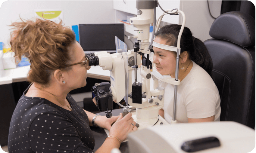
On the day of your appointment, one of our retina specialists will sit with you and discuss your symptoms or concerns, assess your vision and eye pressure. Then you will require eye drops to dilate your pupil. These eye drops take time to work, and once your pupil is dilated your vision will become slightly blurry and sensitive to light. This blurriness may last for a few hours, and then your vision will return to normal as the eye drops wear off naturally.
Macular Assessment Tests
If there are concerns about your macula, you may require some additional specialised tests that focus on the health and function of your macula. These tests include:
1 . Optical Coherence Tomography (OCT) and OCT Angiography
This is a non invasive imaging scan that provides detailed cross sectional images of the macula. It assesses the layers of the macula that cannot be seen on a slit lamp (microscoped examination. It is used to detect and monitor macular and retinal conditions.
2 . Retinal Photography and Auto fluorescence
This is a specialised camera that takes images of your retina to detect and monitor macular and retinal conditions
3 . Fluorescein Angiography / Indocyanine Green Angiography
This is a diagnostic procedure used to examine the blood flow in the retina. It involves injecting a special dye called Fluorescein or ICG into a vein in your arm, then taking a series of photographs as the dye circulates through the blood vessels in the retina. The test identifies areas of leaking or abnormal blood vessels, blockages, swelling or other changes that may affect the health and function of the retina.
At Nexus Eye Care, it is our priority to provide thorough and exceptional eye care to all our patients. After your assessment and tests are complete, your Retina Specialist will sit with you and explain all your results and discuss if any retinal disease treatment is required.